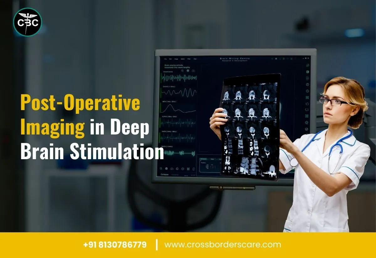
Deep Brain Stimulation (DBS) therapy offers significant results to patients with different movement disorders. It covers dystonia, essential tremor, and Parkinson’s disease. The implantation of the electrodes in the DBS process focuses on modulating the neural activity which relieves the key movement disorder symptoms.
DBS gets the best help from dedicated post-operative imaging confirming its success and long-term effectiveness to patients. Read on to learn more about the impact of the post-operative imaging in Deep Brain Stimulation surgery .
Types of post-operative imaging in Deep Brain Stimulation
The different forms of post-operative imaging in Deep Brain Stimulation covers:
Computed Tomography (CT)
It helps quickly verify the position of the DBS electrodes and find post-surgery acute complications like shifts in electrode position or haemorrhages in brain tissues.
Magnetic Resonance Imaging (MRI)
It offers high-resolution images of the brain and helps in visualising the precise location of the implanted electrodes in the brain. MRI is suitable for regular follow-up and helps in detailed anatomical information of the patient’s brain after DBS therapy.
Positron Emission Tomography (PET) and Single Photon Emission Computed Tomography (SPECT)
The combination of PET and SPECT evaluates the functional impact of DBS on brain activity. It offers quick insights into brain function after DBS therapy and significantly contributes to the cost of Deep Brain Stimulation surgery.
Impact of post-operative imaging in Deep Brain Stimulation
The key factors highlighting the importance of post-operative imaging in Deep Brain Stimulation cover:
Confirmation of the electrode placement
The key benefit of post-operative imaging helps in verifying the position of the electrodes in the DBS. This placement is essential for ensuring that the stimulation is offering desired therapeutic effects while minimising the potential side effects.
The electrode tips and location of the electrodes are quickly determined post-surgery during the follow-up visits. Any misplacement of electrodes calls for quick revision surgery or adjustments in stimulation settings.
Quick identification of complications
The possible DBS complications like mechanical issues with device hardware, infections, or haemorrhages can be identified easily by imaging techniques. Detecting the bleeding within the brain and assessing the integrity of the DBS system is best facilitated by post-operative imaging.
CT scans can help in immediate surgery for treating haemorrhages and MRI can help in assessing the DBS system hardware for possible mechanical failure or infection risks.
Guided programming and adjustments
DBS requires precise programming for optimised therapeutic results. The results from the post-operative imaging help in adjusting the different stimulation parameters according to the electrode placement.
Long-term monitoring
With natural ageing, brain anatomical changes lead to shifts in the DBS system or disease progression. MRI helps in easy tracking of brain structure changes affecting the effectiveness of DBS therapy.
The dedicated scans like MRI help in assessing the shift of electrodes in their original positions making necessary adjustments in the stimulation settings.
Supporting dedicated research and development
The advanced imaging techniques offer the right support in the dedicated research and development of DBS therapies. Functional imaging studies help find the impact of DBS on brain connectivity and activity for improved optimization and understanding of DBS effectiveness.
The different insights from the imaging studies help in the smooth development of the new DBS systems and offer improved surgical techniques. These guided programming and DBS system adjustments help in managing the overall Deep Brain Surgery cost in India.
Ending note
Post-operative imaging in Deep Brain Stimulation is integral for the long-term success of this process. The key benefits of using post-operative imaging include the confirmation of electrode placement, identification of complications, required programming, long-term monitoring, and dedicated research for further improvements.
Cross Border ensures the best patient care, advanced diagnostics, and high-quality treatment in the best state-of-the-art hospitals.
September 9, 2024


