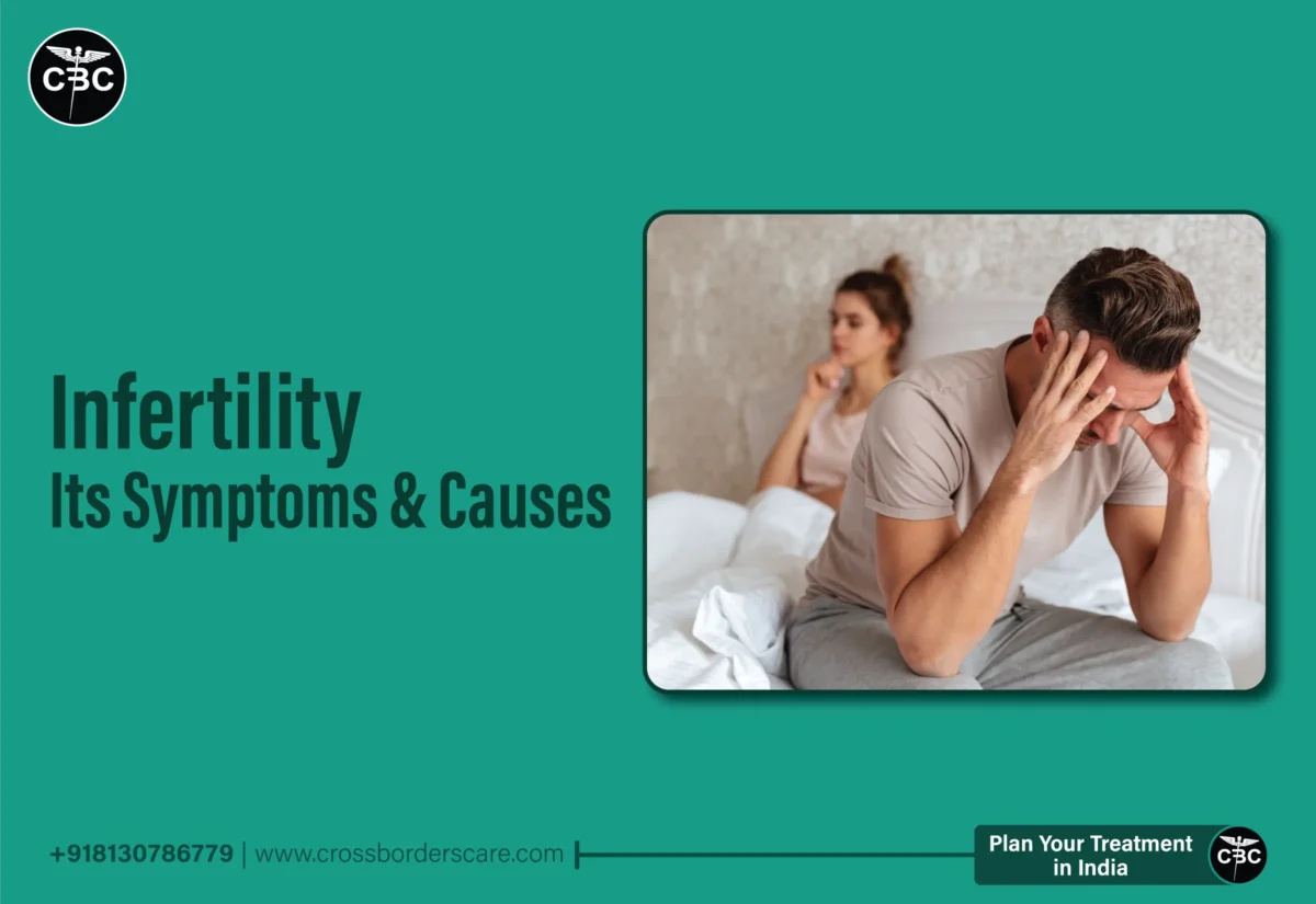
Categories
Uncategorized

Bentall Procedure: Preparation, Recovery and Long-Term Care

The Bentall surgery, commonly referred to as the Bentall procedure, is a sophisticated cardiac surgical treatment used to address a variety of diseases that affect the aorta and aortic valve. It includes synthetic or biological replacements for the aortic root, aortic valve, and ascending aorta. This procedure is usually used to treat aortic aneurysms and aortic valve problems simultaneously. This technique successfully treats both the aortic root and valve problems, increasing overall heart function and lowering the risk of complications related to aortic disorders. The choice of valve and graft material is made depending on the patient’s health and the surgeon’s preference. Bentall surgery in India is an entirely safe procedure that has helped thousands of patients.

The procedure of the Bentall Surgery

It involves replacing the aortic root, the aortic valve, and the ascending aorta. The steps of the Bentall surgery are described below:

● Anesthesia: The patient is put under general anesthesia before surgery. This guarantees that the patient will be asleep and pain-free during the procedure.

● Make an Incision: The surgeon makes A significant incision in the chest, usually via a median sternotomy. To access the heart and aorta, one must cut through the sternum, the bone that forms the breastbone. This opens up the heart and gives a clear glimpse of it.

● Cardiopulmonary Bypass (CPB): The patient is connected to a heart-lung machine (CPB) to complete the procedure. The heart can be operated on without the requirement for blood circulation since this device temporarily takes over the activities of the heart and lungs.

● Aortic Root Removal: The surgeon removes the damaged segment of the aorta, including the aortic valve and any weakening or aneurysmal tissue. This usually involves dividing the coronary arteries and removing the aortic valve.

● Replacing the aortic valve: A prosthetic valve is inserted to replace the removed aortic valve during aortic valve replacement. Mechanical (made of metal) or biological (often from a pig or cow, medicated to prevent rejection) valves are also options. The replacement valve is firmly stitched into position.

● Aortic Graft Placement: The excised aortic root and ascending aorta are replaced with a synthetic tube (graft), frequently composed of materials like Dacron. The new prosthetic valve is attached to this graft by sewing it in place. The previously separated coronary arteries are re-implanted into this transplant to ensure they receive blood flow.
● Closure: After ensuring the prosthetic valve and graft are securely in place, and blood usually flows via the coronary arteries, the surgeon sews up the chest incision. The sternum may need to be tightly closed using wires or other surgical supplies.

● Weaning from CPB: When a patient has weaned off a heart-lung machine, their heart can start pumping blood again.

● Monitoring and Recovery: Following surgery, the patient is continuously watched in the intensive care unit (ICU) to ensure steady recovery.

Preparation

Preparing for a Bentall procedure is essential to ensure the patient’s safety and the operation’s success. Here’s a detailed explanation of the process:

● Medical Evaluation: The patient receives a thorough evaluation before a Bentall treatment. This includes a comprehensive review of one’s medical history and diagnostic procedures like echocardiograms, CT scans, and angiograms. These tests aim to decide whether the patient is a good candidate for surgery.

● Discussion with the Surgical Team: The patient meets with the surgical team, which includes the anesthesiologist and the cardiac surgeon, to discuss the surgery. The patient can clear any doubts, ask questions, and learn about the advantages and disadvantages of the procedure.

● Medication Modification: Before surgery, the patient’s medications may need to be modified or temporarily halted. This includes medicines that can affect anesthesia, anticoagulants, antiplatelet drugs, etc.

● Stop smoking: Patients who smoke are strongly urged to give up before surgery since it might hinder wound healing and raise the possibility of problems.

● Dental Checkup: Dental health is crucial before heart surgery since dental infections can spread to the valves. Patients might need to get their teeth checked and treated if necessary.

● Blood transfusion: If a blood transfusion is planned during or after the Bentall procedure, patients may occasionally donate their blood before surgery.

● Fasting: The patient is told to abstain from food or drink for a certain amount before surgery, usually the night before. This lessens the possibility of aspiration.

● Final Preoperative Check: On the day of the operation, the surgical team does a last preoperative check to validate the patient’s identity, confirm the surgical procedure, and ensure all necessary tools and supplies are available.

Recovery

Here are some recovery tips to consider after undergoing a Bentall surgery in India

● Hospital Stay: Depending on the patient’s health and the surgical strategy chosen, the initial recovery phase often entails a hospital stay of seven to ten days.

● Pain management: After major cardiac surgery, pain and discomfort are typical. The medical staff will administer the proper pain management, which may entail using drugs or other pain reduction methods. It is crucial to let the medical staff know how much pain you are experiencing so that they can modify your pain management strategy accordingly.

● Monitoring: Once your health has stabilised, you will be closely watched in a regular hospital rather than the cardiac critical care unit (ICU). Heartbeat, vital signs, and other factors will be carefully monitored.

● Medication: You will be given prescriptions for drugs to help you manage different elements of your rehabilitation, including pain, blood thinners to avoid clots, and medicines to help your heart operate. Follow the directions on all prescriptions.

● Wound Care: It’s essential to take care of your surgical incision. Follow the healthcare team’s recommendations for dressing changes and wound care. Keep the incision clean and dry.

● Cardiac Rehabilitation: Your healthcare provider might advise enrolling in a cardiac rehabilitation program after discharge. Through exercise, education, and dietary modifications, this structured program aids in improving your cardiovascular health.

Long-Term Care

Here are some tips on how to take long-term care after the procedure:

● Attendance at Follow-up Appointments: Show up for all scheduled follow-up consultations with your cardiologist and heart surgeon. These consultations are crucial for keeping track of your development and modifying your treatment strategy.
● Heart-Healthy Diet: Consume a heart-healthy diet low in sodium, cholesterol, and saturated and trans fats. Consume a lot of fruits, vegetables, whole grains, lean proteins, and omega-3 fatty acid-rich meals.
● Frequent Exercise: Follow your healthcare team’s recommendations and exercise regularly. Exercise helps maintain weight, enhance overall fitness, and improve cardiovascular health. Follow your doctor’s advice at all times, but begin with easy exercises and work up to more rigorous ones.
● Alcohol consumption: Limit alcohol consumption to reasonable levels because excessive alcohol can damage the heart. The same goes for caffeine. Caffeine should be consumed with caution because it can sometimes disrupt heart rhythm.
● Stress management: Use relaxation methods, mindfulness, meditation, or counselling to manage stress healthily. High levels of stress can significantly impact heart health.

As a result, Bentall surgery in India restores health and vitality to individuals with serious heart conditions by addressing aortic root and valve issues.

Categories
Uncategorized

Prostatectomy: Purpose, Types, Procedure, Risks and Recovery

A prostatectomy can surgically remove the prostate gland. This can be done for various reasons, such as managing specific benign prostatic diseases or treating prostate cancer. There are many types of prostatectomies, including transurethral resection of the prostate (TURP), which removes a portion of the prostate through the urethra, and radical Prostatectomy, which removes the entire prostate gland. The exact medical issue and the severity of the case determine the treatment to be used. Prostatectomy surgery in India is a safe and effective procedure for patients who suffer from prostate-related conditions. A healthcare expert should be consulted for detailed information and counseling.

Purpose

A prostatectomy is primarily performed to treat diseases associated with the prostate gland. The patient’s condition and unique circumstances help to determine the type of Prostatectomy and surgical strategy that will be performed. Depending on the underlying ailment, this surgical technique may have several objectives:

  • Treating prostate cancer: Treatment for prostate cancer is one of the most frequent uses of a prostatectomy. If the cancer is limited and hasn’t migrated to other parts of the body, the aim is to remove the malignant prostate tissue in this scenario.
  • Benign Prostatic Hyperplasia (BPH): A prostatectomy may be performed to remove extra prostate tissue that is obstructing the urethra and causing urinary issues in cases of benign prostatic hyperplasia. In benign prostatic hyperplasia, an enlarged prostate can cause urine symptoms. This primarily aims to alleviate urinary problems.
  • Prostatitis: In extremely rare instances of severe and persistent prostatitis (prostate inflammation), a prostatectomy may be an option as a last resort to relieve symptoms that haven’t improved with other therapies.

Types

The patient’s medical condition, the size of the prostate, the existence of cancer, and the surgeon’s experience all play an important role in choosing the type of Prostatectomy that is performed. The patient must consult a healthcare professional before undergoing the procedure. Here are the main types of Prostatectomy:

  • Radical Prostatectomy: The most popular form of Prostatectomy for the treatment of prostate cancer is radical Prostatectomy. The entire prostate gland, as well as any adjacent lymph nodes, must be removed. Different methods can be used to perform a radical prostatectomy:
  • Open surgery: A significant abdominal incision is made to access the prostate.
  • Laparoscopic Prostatectomy: A minimally invasive procedure in which the surgeon is guided via a camera through small incisions.
  • Robotic-assisted laparoscopic Prostatectomy: A laparoscopic surgery in which the surgeon is helped by a robotic technology to carry out the process more precisely.
  • Robotic-assisted laparoscopic Prostatectomy: A laparoscopic surgery in which the surgeon is helped by a robotic technology to carry out the process more precisely.
  • Transurethral resection of the prostate (TURP): Benign prostatic hyperplasia (BPH) is treated with the transurethral resection of the prostate (TURP). The extra prostate tissue blocking the urethra is removed rather than the complete prostate. The urethra usually does this procedure through specialized devices.
  • Transurethral prostate incision (TUIP): Similar to TURP, transurethral prostate incision (TUIP) is used to treat BPH. To treat urinary problems, tiny incisions are made in the prostate gland. It is typically used for BPH situations that are less severe.
  • Simple Prostatectomy: This surgery is utilized when TURP or TUIP is ineffective for treating very big prostates. It entails keeping the prostate’s outer shell intact while just removing the inner portion of the prostate that is obstructing the urinary tract.
  • Laser techniques: Holmium laser enucleation of the prostate (HoLEP) and GreenLight laser therapy are two examples of laser techniques that can be used to treat BPH. These methods reduce urinary problems by vaporizing or removing extra prostate tissue with laser radiation.

Procedure

Whether it’s a radical prostatectomy for cancer or a less invasive procedure for benign disorders like BPH, the prostatectomy procedure may vary depending on the type of surgery being done. The most common method of Prostatectomy involves the following fundamental steps:

  • Preparation: The patient will normally undergo several medical examinations and tests before surgery to determine their general health and fitness for the procedure. They might also need to change their medication and adhere to certain dietary requirements.
  • Anesthesia: The patient receives either regional anesthesia, which numbs the lower body, including the surgical area, or general anesthesia, which puts them into a deep sleep.
  • Incision: An incision will be made in the abdomen or the lower section, depending on the surgical method (open, laparoscopic, or robotic-assisted). Small incisions are made to allow specialized equipment and a camera during robotic or laparoscopic treatments.
  • Prostate Removal: The surgeon meticulously dissects and removes the entire prostate gland and any potentially cancerous lymph nodes or surrounding tissues. All malignant tissue must be removed, but surrounding tissues, including blood vessels and nerves necessary for sexual and urine function, must be preserved.
  • Reconstruction: The surgeon may repair the urinary system after removing the prostate. To restore urine function, the bladder may need to be sutured back to the urethra.
  • Closure: The incisions are stitched or stapled shut by the surgeon, and the surgical site is dressed or bandaged.
  • Recovery: Patients who emerge from anesthesia are closely watched in a recovery area. The time that a patient stays in the hospital for recovery will depend on the type of surgery they underwent and their health. Everybody recovers differently following a prostatectomy, but most people need time to relax and heal.

Risks

Like any other surgical procedure, prostatectomies have possible risks. The risks may vary depending on the type of Prostatectomy performed and the patient’s health. The following are some typical dangers of Prostatectomy:

  • Bleeding: Both during and after surgery, bleeding can occur. Additional surgical operations can be required in some cases to stop bleeding.
  • Infection: The urinary tract and the surgical site are at risk of contracting an infection. Antibiotics are frequently effective in treating this, although other treatments can also be used.
  • Blood Clots: Patients undergoing surgery risk developing blood clots, which may spread to other parts of the body or the lungs.
  • Urine Incontinence: Some men who have undergone Prostatectomy may have urine incontinence, which can range from little leakage to total loss of urinary control, either temporarily or permanently.
  • Erectile Dysfunction: After a prostatectomy, men may develop temporary or permanent erectile dysfunction depending on the surgical strategy and the level of nerve-sparing treatments.
  • Damage to Surrounding Structures: During surgery, it’s possible to unintentionally harm nearby structures like the rectum, blood arteries, or nerves.
  • Complications of anesthesia: Anesthesia has its dangers, such as unfavorable reactions, respiratory issues, or allergies.
  • Recurrence: Even after a successful prostatectomy, prostate cancer patients still have the chance of a cancer relapse. This can call for additional care, such as radiation or hormone therapy.

Recovery

Recovery time after prostatectomy can vary from person to person. Here is some general advice on healing that can be useful:

  • Follow Medical Advice: Comply with any prescription directions, wound care instructions, and other post-operative advice from your surgeon and medical staff.
  • Activity and Rest: While taking it easy and letting your body heal is vital, doing some light exercise is also crucial. Walking is frequently advised to enhance circulation and prevent blood clots.
  • Pain Control: If you’re in too much pain, take your painkillers as directed and let your medical staff know.
  • Diet: Comply with any dietary recommendations made by your medical experts. Healing can be aided by drinking plenty of water and eating a healthy diet.
  • Exercises for the Pelvic Floor: Kegel exercises for the pelvic floor, also known as pelvic floor exercises, can aid with bladder control. Ask the doctor to explain them in detail.
  • Avoid Heavy Lifting: To prevent strain on the surgical area, refrain from heavy lifting or hard activity for a few weeks or as directed by your surgeon.
  • Take care of the incision: Maintain the surgical incision clean and dry. Change dressings according to instructions, and report any infection-related symptoms such as edema, discharge, or increasing redness.

For prostate-related conditions, prostatectomy surgery in India offers cutting-edge solutions. With skilled surgeons and state-of-the-art facilities, patients can get effective treatments here.

Categories
Uncategorized

Anterior Cervical Discectomy and Fusion – Indications and Outcomes

Anterior cervical discectomy and fusion (ACDF) is a surgical treatment used to treat neck pain and neurological problems caused by degenerative cervical discs in the cervical spine. In an ACDF procedure, an orthopedic or neurosurgeon gains access to the cervical spine from the front of the neck, removes the herniated or slipped disc or discs with care, and then replaces them with a bone graft or artificial spacer.

This spinal segment is stabilized through fusion, which stops additional movement and relieves discomfort. Herniated discs, degenerative disc disease, and cervical spondylosis are among the disorders that frequently require the ACDF operation. Although it does restrict some neck movement because of the fused segment, it frequently leads to enhanced neck function and a decrease in accompanying complaints. There could be hazards with surgery, as there always are. And as with any surgery, there are potential risks and benefits that should be discussed with a healthcare professional before the procedure. Also, Anterior Cervical Discectomy and Fusion (ACDF) cost in India can vary significantly depending on factors such as the hospital, location, and the specific requirements of the procedure.

Surgical Indications

Anterior cervical discectomy and fusion (ACDF) is recommended for a number of cervical spine issues that cause pain, neurological symptoms, or spinal instability. The following are typical surgical indications for ACDF:

  • Herniated Disc: When a herniated cervical disc pushes on spinal nerves or the spinal cord and results in symptoms like neck pain, arm pain, numbness, weakness, or tingling, ACDF is frequently advised.
  • Degenerative Disc Disease: ACDF may be considered to treat symptoms and stabilize the spine when cervical discs gradually deteriorate, causing chronic neck pain, radiculopathy (nerve compression), or myelopathy (compression of the spinal cord).
  • Cervical Spinal Stenosis: Narrowing of the spinal canal in the cervical region is known as cervical spinal stenosis. If this condition exists, ACDF can assist relieve spinal cord compression, which can cause weakness, balance problems, and other neurological concerns.
  • Cervical Spondylosis: This age-related disorder affects the neck’s joints and spinal discs by causing wear and strain. When spondylosis causes discomfort, nerve compression, or instability, ACDF may be used.
  • Trauma or Cervical Fractures: In situations of cervical spine fractures, particularly those that result in instability or endanger the integrity of the spinal cord, ACDF may be required to regain stability and stop additional damage.
  • Failed Conservative Treatment: ACDF may be explored as a surgical option when conservative therapies like physical therapy, medicine, and rest fail to relieve cervical spine symptoms.
  • Tumors: Using ACDF, cervical spine tumors or infections that have spread to the spinal discs or other tissues can be removed.

Due to competitive Anterior Cervical Discectomy and Fusion (ACDF) costs in India patients often choose to undergo ACDF in India.

Procedure

Anterior cervical discectomy and fusion is a surgical treatment used to treat a variety of cervical spine disorders. Here is an overview of the procedures:

  • Anesthesia: To ensure that the patient is unconscious and pain-free throughout the treatment, general anesthesia is often administered before surgery.
  • Positioning the patient: The patient is lying on their back, and the area around their neck is exposed and cleansed. Usually on the right or left side, a tiny incision is made at the front of the neck.
  • Access to the Spine: The surgeon carefully separates and moves aside the soft tissues, such as muscles and blood vessels, to access the cervical spine. Direct access to the afflicted area is provided by this method from the front of the neck.
  • Removal of the Disc: The herniated or injured disc is then carefully removed. To increase precision, surgical tools like microscopes or surgical loupes may be employed.
  • Placement of a Spacer or Bone Graft: A spacer or bone graft is put into the vacant disc area to preserve the correct spacing between adjacent vertebrae and to encourage fusion. The transplant may be either an allograft (donor bone) or an autograft (bone taken from the patient’s own body, commonly the hip).
  • Fixation: To stabilize the spine and hold the vertebrae in place, the surgeon frequently uses hardware like metal plates, screws, or cages. The fusion process is supported by this hardware.
  • Closing the Incision: The surgeon uses sutures or staples to close the incision after making sure the graft and hardware are properly positioned.
  • Recovery: After being observed in the recovery area, the patient is sent to a hospital room for observation and post-operative care.

The affordable Anterior Cervical Discectomy and Fusion (ACDF) cost in India makes it an attractive option for both domestic and international patients seeking spinal surgery.

Post Operative Precautions

To guarantee a successful recovery and reduce problems after anterior cervical discectomy and fusion (ACDF) surgery, post-operative precautions are essential. Here are some general post-operative safety measures to take into account:

  • Neck Immobilization: You might be forced to wear a neck brace or collar to prevent your neck from moving and to give support. Pay attention to your surgeon’s recommendations for when and how to wear it.
  • Restricted activities: You must first refrain from engaging in any activity that could put stress on your neck or spine. Heavy lifting, bending over at the waist, and demanding activities fall within this category. Guidelines on when it is safe to gradually resume these activities will be given by your surgeon.
  • Medication: Follow the directions on the prescription for antibiotics and painkillers. These drugs can aid in pain management and infection prevention.
  • Proper care of incision: Maintain the surgical incision by keeping it clean and dry. Report any signs of infection, such as redness, swelling, or discharge, and adhere to any wound-care recommendations your surgeon may have given you.
  • Avoid drinking and smoking: If you smoke, it’s best to stop because it can slow down the healing process. Limit your alcohol intake as well because it may affect your ability to recover and take your medication.
  • Follow-Up Consultations: Keep all of your surgeon’s scheduled follow-up appointments. These consultations are necessary for tracking your development and correcting any issues.
  • Physical Therapy: Participate in physical therapy sessions as directed to gradually increase neck strength and mobility.
  • Driving Restrictions: Because of the neck brace and painkillers, you might not be able to drive for a certain time after surgery.

For people suffering from cervical spine conditions that cause pain and neurological symptoms, ACDF is a great surgical option. A successful ACDF depends not just on the surgeon’s skill, but also on the patient’s commitment to post-op care and rehabilitation. Patients can look forward to a brighter, pain-free future by working closely with healthcare professionals. Anterior Cervical Discectomy and Fusion (ACDF) cost  in India is typically lower than in many Western countries, it’s essential for patients to thoroughly research healthcare providers to ensure they receive safe and effective treatment.


Categories
Uncategorized

Lung Cancer – Types, Symptoms, Diagnosis and Treatment

Lung cancer is a deadly illness caused by the uncontrolled growth of cells in the lung tissues. Millions of people die from it worldwide, making it one of the most prevalent types of cancer. Long-term exposure to carcinogens such as radon gas, environmental contaminants, and cigarette smoke usually leads to the development of lung cancer. Small cell lung cancer and non-small cell lung cancer are the two main forms of lung cancer, each with distinctive characteristics and therapeutic choices.  Lung cancer symptoms include fatigue, weight loss, shortness of breath, chest pain, and chronic coughing. Successful lung cancer treatment depends heavily on early detection. Robotic lung cancer treatment in India is a popular and effective treatment method.

Types of Lung Cancer

There are two main types of lung cancer: non-small cell lung cancer and small cell lung cancer.

Non-Small Cell Lung Cancer (NSCLC): Making up nearly 85% of all occurrences of lung cancer, NSCLC is the most prevalent kind. It is further broken down into various types of subtypes:

  • adenocarcinoma: The most prevalent subtype, adenocarcinoma, frequently affects non-smokers or former smokers. It typically begins in the lungs’ outermost regions and can migrate to the lymph nodes.
  • Squamous Cell Carcinoma: This subtype frequently appears in the bronchial tube lining. It may result in symptoms like coughing and shortness of breath and is frequently linked to a history of smoking.
  • Large Cell Carcinoma: This subtype is less frequent and has the tendency to advance rapidly. It can occur anywhere in the lung and is frequently considered to have a dismal prognosis.

Depending on the cancer’s subtype and stage, NSCLC treatment options include surgery, radiation therapy, chemotherapy, targeted treatments, and immunotherapy.

Small cell lung cancer (SCLC): The less frequent but more dangerous kind of lung cancer is known as small cell lung cancer (SCLC). It frequently spreads and grows quickly. The two stages of SCLC are limited, which affects only one lung, and extensive, which affects more than one lung. They are significantly linked to cigarette use.

SCLC is frequently treated with chemotherapy and radiation therapy due to its aggressive nature. Since cancer has typically spread by the time of detection, surgery is rarely an option. Robotic lung cancer treatment in India can also be used for this.

Stages of Lung Cancer

A system called lung cancer staging is used to describe how far the disease has gone throughout the body. It aids medical professionals in selecting the best course of treatment. Each stage has specific treatment options, like robotic lung cancer treatment in India, with earlier stages often having a better prognosis and a wider range of treatment options.

  • Stage 0: Also referred to as carcinoma in situ, this stage denotes the presence of abnormal cells exclusively in the lining of the airways and excludes the invasion of deeper lung tissues.
  • Stage I: The cancer has not progressed to the lymph nodes or any distant sites and is contained to the lung.
  • Stage II: Though not at distant sites, the tumor may have gotten bigger and spread to surrounding lymph nodes.
  • Stage III: In this stage, the cancer has spread to the lymph nodes close by and may even have infiltrated surrounding tissues like the diaphragm or chest wall. Stage III is further broken down into sub-stages, where Stage IIIA denotes lymph node involvement on the same side of the chest and Stage IIIB denotes lymph node involvement on the other side.
  • Stage IV: At this point, the cancer has spread to distant organs such as the brain, liver, bones, or other sections of the body. This lung cancer is regarded as progressed or metastatic.

Symptoms

The type of lung cancer, its stage, and individual circumstances can all affect the symptoms of lung cancer. Lung cancer patients’ prognoses can be considerably enhanced by early detection and therapy. An efficient way for treating lung cancer is robotic lung cancer treatment in India. Typical signs include:

  • Persistent cough: A persistent cough is frequently an early indicator of lung cancer. It either doesn’t go away or gets worse over time.
  • Breathlessness: Breathlessness and breathing difficulties can result from lung malignancies that obstruct or constrict airways.
  • Chest Pain: Chest pain or discomfort brought on by lung cancer may get worse when you cough, laugh, or take heavy breaths.
  • Unexpected weight loss: A number of problems, including lung cancer, might be indicated by sudden and unexplained weight loss.
  • Fatigue: Even with little exercise, feeling exceedingly exhausted or weak is a common symptom.
  • Wheezing: Narrowed or obstructed airways can cause a whistling or wheezing sound when breathing.
  • Hoarseness: Lung tumors that are pushing on the nerves or airways can cause vocal changes, such as hoarseness.
  • Frequent Infections: Lung cancer patients may have weakened immune systems, which makes them more vulnerable to respiratory infections like pneumonia and bronchitis.
  • Coughing Up Blood: When lung cancer is more advanced, coughing up blood or bloody mucus might be a sign.
  • Loss of Appetite: As cancer gets worse, many people experience a loss of appetite and accidental weight loss.
  • Pain in bones or spine: The spine, ribs, or other bones may experience bone pain if the malignancy has spread to them.

Diagnosis

The diagnosis of lung cancer involves several steps to accurately determine the disease’s presence, type, and stage. It typically begins with a thorough medical history and physical examination. If lung cancer is suspected, various imaging tests such as chest X-rays, CT scans, and PET scans are conducted to visualize the lungs and identify any abnormalities or tumors.

  • A definitive diagnosis usually requires obtaining a tissue sample for examination. This can be done through procedures like bronchoscopy or a needle biopsy. Sometimes, a surgical biopsy may be necessary to extract a larger tissue sample for examination.
  • Once a tissue sample is obtained, a pathologist analyzes the cells under a microscope to determine if they are cancerous and, if so, what type of lung cancer it is.
  • To assess the extent of the disease, staging is conducted to determine if the cancer has spread beyond the lungs and to what extent. Staging helps healthcare professionals to determine the best treatment approach. Lung cancer is categorized into stages ranging from I to IV, with higher stages indicating more advanced disease. Modern techniques like robotic lung cancer treatment in India can also be used.

Treatment

The type of lung cancer, its stage, the patient’s general health, and personal preferences are only a few of the variables that affect how the disease is treated. Modern approaches like robotic lung cancer treatment in India are becoming increasingly popular. Treatment strategies comprise:

  • Surgery: During surgery, the tumor and adjacent lymph nodes are removed. It works well for non-small cell lung cancer in its earliest stages.
  • Radiation therapy: High-energy rays are used in radiation therapy to target and kill cancer cells. It can be combined with other treatments for advanced stages of lung cancer or used as the main treatment for early-stage lung cancer.
  • Chemotherapy: This treatment uses medication to either kill or slow the growth of cancer cells. It can be administered prior to surgery to reduce tumor size, following surgery to eliminate cancerous cells still present, or for advanced lung cancer to manage symptoms.
  • Targeted Therapy: Drugs that specifically target genetic abnormalities or proteins that fuel the proliferation of cancer cells are known as targeted treatments.
  • Immunotherapy: Immunotherapy works by enhancing the immune system’s capacity to identify and combat cancer cells.
  • precision medicine: Using precision medicine, treatment is customized according to the unique genetic composition of a patient’s tumor. It can assist in locating more effective, individualized therapy solutions.
Categories
Uncategorized

Prostatectomy: Purpose, Types, Procedure, Risk and Recovery

A prostatectomy is a medical surgery to remove the prostate gland, an organ about the size of a walnut found in males right below the bladder. This procedure is frequently used to treat benign prostatic hyperplasia (BPH), a non-cancerous growth of the prostate that can cause urinary issues and prostate cancer. There are various ways to do a prostatectomy, each with advantages and disadvantages, such as open surgery, laparoscopic methods, and robot-assisted treatments.

The particular state of the patient and the surgeon’s experience frequently influence the procedure selection. While a prostatectomy can effectively treat specific health problems, it can also have unfavorable outcomes like erectile dysfunction and urine incontinence. Therefore, cautious thought and consultation with a medical expert are crucial for patients undergoing this procedure.

Purpose

Prostate cancer is one of the most prevalent cancers in men. A prostatectomy can be done to remove the cancerous tissue when the cancer is contained to the prostate gland and hasn’t spread to other parts of the body. It is frequently undertaken when other therapies like radiation therapy or active surveillance are not preferred. Benign prostatic hyperplasia (BPH), a disorder marked by an enlarged prostate, can also be treated with a prostatectomy to relieve symptoms. The urinary function and quality of life can be improved by removing part or all of the prostate when BPH substantially obstructs the urinary tract.

Types

There are various types of prostatectomy operations, which involve surgically removing the prostate gland. The patient’s condition, the prostate’s size, and the surgeon’s experience all play a role in determining which operation is best. Here are the main categories:

  • Open retropubic prostatectomy: In the conventional “open retropubic prostatectomy method,” the surgeon creates a cut in the lower abdomen to reach and remove the prostate gland. It gives the surgeon easy access and visibility but often requires extended recuperation.
  • Open perineal prostatectomy: An incision is made between the anus and the scrotum to access the prostate during an open perineal prostatectomy. Compared to the retropubic method, it is less frequent but may be preferred in some circumstances.
  • Laparoscopic Prostatectomy: This minimally invasive process uses specialized tools and tiny incisions. It can be performed as a robot-assisted or laparoscopic radical prostatectomy. The surgeon’s precision and skill are improved with the robotic version.
  • Benign prostatic hyperplasia (BPH) is treated with the transurethral resection of the prostate (TURP). Extra prostate tissue is removed using a specialized tool introduced through the urethra. It is frequently used for less severe cases of BPH since it is less intrusive than other types of prostatectomy.
  • Robotic-assisted radical prostatectomy: The physician uses A robotic system during a robotic-assisted radical prostatectomy to complete the process through small incisions. The robot provides greater control and better precision.

Procedure

The precise steps and techniques may change depending on the type of prostatectomy being performed, such as open surgery, laparoscopic surgery, or a robotic-assisted approach. A general description of the process is given below:

  • Preparation: To ensure the patient is in good health for the procedure, they will typically undergo several preoperative examinations and evaluations. Blood testing, imaging tests, and discussions about any allergies may be part of this.
  • Anesthesia: The patient is unconscious and has no pain perception during the surgery because it is done under general anesthesia.
  • Incision: The size and position of the incision will depend on the type of prostatectomy
  • Prostate gland access: The surgeon gently cuts through the tissue layers to reach the prostate gland. Specialized tools and a camera are used in robotic or laparoscopic procedures to view and access the area.
  • Surgery to Remove the Prostate: The surgeon akes out the prostate. Although the precise method can vary, it typically entails severing and closing blood ducts and veins that link to the prostate to enable its removal.
  • Lymph node examination: Prostate cancer patients have their adjacent lymph nodes removed to see if the cancer has spread may have this procedure.
  • Closure: After the prostate is removed, the surgeon skillfully stitches or stapled shut the wounds. To remove extra fluid, a surgical drain may occasionally be placed.
  • Recovery and Postoperative Care: Patients who emerge from anesthesia are closely watched in a recovery area.
  • Follow-up: Patients will schedule routine follow-up sessions with their medical team to assess their progress, manage any side effects, and review additional treatment options.

Risks Associated

Various factors can affect the risks, including the patient’s general health, the type of prostatectomy used, and the surgeon’s expertise. The following are some typical dangers and issues related to prostatectomy:

  • Infection: The urinary tract and the surgical site are at risk of contracting a disease. Medicines and careful surgical methods are used to manage this danger.
  • Bleeding: Bleeding can happen both during and following surgery. Even while surgeons take measures to prevent bleeding, it occasionally requires further treatments.
  • Blood Clots: Having surgery increases the chance that blood clots will develop in the legs or will spread to the lungs. This risk is decreased by using strategies like early mobilization and blood-thinning drugs.
  • Urinary incontinence: Some men may develop brief or chronic urinary incontinence after a prostatectomy.
  • Erectile Dysfunction: Prostatectomy procedures can potentially cause erectile dysfunction, particularly when the erection-related nerves are damaged.
  • Lymphedema: Prostate cancer patients who have their lymph nodes removed may develop lymphedema or swell brought on by a disturbance of the lymphatic system.
  • Anesthesia-related problems: General anesthesia has several concerns, including adverse drug responses and respiratory issues.
  • Damage to Neighboring Structures: In rare circumstances, unintended damage to surrounding structures during surgery, such as the rectum, bladder, or blood arteries, may necessitate additional treatments.

Recovery

  • Medical Advice: Always follow the advice of your doctor and the rest of the medical staff. This includes following food and exercise advice, taking prescribed medications, and attending follow-up appointments.
  • Observe the surgical site: Look for infection-related symptoms like fever, redness, edema, excessive drainage, or swelling at the location of the incision. Inform your healthcare professional as soon as you have any unsettling symptoms.
  • Pain management: Following surgery, you can feel pain or discomfort. Take your painkillers as directed, and let your healthcare staff know if your pain is not under control.
  • Proper Diet: Follow any dietary advice given to you by your medical staff. You might begin with a liquid or soft diet in the days immediately following surgery and then gradually introduce your regular food.
  • Rest and Activity: As your surgeon directs, gradually increase your activity level. It is frequently advised to walk to aid healing and prevent blood clots. You refrain from intense activities and heavy lifting until your doctor gives the all-clear.
  • Support on an emotional level: Recovering emotionally might be complex. Ask friends, family, or support groups for assistance.
  • Avoid Smoking and Drinking Too Much: Smoking and drinking too much alcohol might slow healing. Attempt to limit or avoid these activities while you’re recovering.
Categories
Uncategorized

Types of Spine Surgery: How to Know You Need One

Spine surgery is a miraculous medical advancement focusing on diagnosing and treating various disorders and conditions affecting the spine. As the central pillar of the support structure of the human body, the spine plays an important part in maintaining posture, mobility, and overall functionality.

Spine surgery involves a wide range of procedures that aim to relieve pain, improve spinal stability, and enhance patients’ quality of life. There are a lot of methods by which surgeons carry out spine surgery, from minimally invasive techniques to complex methods like robotic spine surgery.

In this article, we will learn more about the methods and advancements within the field of spine surgery that continue to evolve and provide relief for those suffering from spinal ailments.

Different Types of Spine Surgery

There are various types of spine surgery, such as robotic spine surgery, artificial disc replacement, decompression surgery, etc. Each surgery aims to address specific spinal conditions and disorders. It is important to note that the type of spine surgery carried out depends on many factors, such as the problem’s severity and the patient’s overall health. Discussing everything with a qualified spine surgeon is important to determine the best surgical approach for each case. Here are some common types:

Decompression surgery: This type of surgery aims to relieve the pressure on spinal nerves by removing portions of bone, herniated discs, or other tissues that may be pressing the nerves. Decompression surgery is carried out to treat problems such as spinal stenosis, herniated discs, and foraminal stenosis.

Spinal fusion: Spinal fusion involves joining two or more vertebrae together using bone grafts, screws, rods, or cages. This surgery aims to stabilize the spine and reduce pain caused by spinal instability, fractures, degenerative disc disease, or certain spinal deformities.

Discectomy: A discectomy involves the removal of some part or all of a damaged or herniated disc in the spine. This procedure helps to relieve pressure on nerves. It can be performed through minimally invasive techniques or traditional open surgery.

Laminectomy involves removing the lamina to widen the spinal canal and reduce pressure on the spinal cord or nerves. The lamina is a part of the vertebra. Laminectomy is often performed to treat spinal stenosis.

Artificial disc replacement: In this procedure, a damaged or degenerated disc is replaced with an artificial disc. The main aim of the disc is to preserve motion and flexibility in the spine. It is an alternative to traditional fusion surgery for certain patients.

Vertebroplasty and kyphoplasty are minimally invasive procedures used to treat vertebral compression fractures caused by osteoporosis or trauma. They involve injecting bone cement into the fractured vertebra to stabilize and relieve pain.

Robotic Spine Surgery: In robotic spine surgery, a surgeon carries out the surgery with the help of a robotic arm. This specialized equipment helps the surgeon navigate and carry out the surgery more efficiently.

Scoliosis surgery: Scoliosis is characterized by abnormal sideways curvature of the spine. Surgery for scoliosis usually involves spinal fusion using rods, screws, and hooks to correct the curvature and stabilize the spine.

Microdiscectomy: This is a minimally invasive version of a discectomy, which uses smaller incisions and specialized instruments to remove a herniated disc with less disruption to surrounding tissues.

How to know when you need spine surgery?

Determining whether you need spine surgery is a complex decision that should be made in consultation with a spine specialist or a neurosurgeon. They discuss surgery’s potential risks and benefits and inform you about alternative treatment options, such as robotic spine surgery. This will help you make a better decision about your spine surgery. Several factors contribute to the decision-making process. It is important to note that spine surgery is carried out only after a thorough evaluation. This includes a detailed evaluation of medical history, physical examination, and imaging studies.

Several factors contribute to the decision-making process. Here are some key indicators that may suggest you need spine surgery:

Persistent and Severe Pain: If you have been experiencing severe back or neck pain for a long time and it does not get better with conservative treatments such as rest, physical therapy, medication, or injections, surgery must be carried out. Severe pain adversely affecting your daily activities and quality of life could indicate surgery.

Neurological Symptoms: Symptoms like numbness, weakness, or tingling in your arms or legs could indicate nerve compression or damage. If these symptoms persist for a long time or worsen despite non-surgical treatments, surgery may be required to relieve the pressure on the affected nerves.

Progressive Neurological Deficits: If you notice a progressive loss of muscle strength, coordination, or bowel/bladder function, it could be a sign of a serious spinal condition. Such a condition may require immediate surgical evaluation.

Failed Conservative Treatments: When conservative treatments such as physical therapy, medications, or injections have been used but there is no significant improvement in the condition, then robotic spine surgery might be considered as the next step.

Structural Spine Issues: Structural problems like spinal instability, spondylolisthesis (slipped vertebra), or spinal deformities like scoliosis or kyphosis may require surgery. Surgery will help to stabilize the spine and prevent further complications.

Traumatic Injuries: In cases of severe spinal injuries caused by accidents or trauma, emergency spine surgery may be required to stabilize the spine and prevent further damage.

Degenerative Disc Disease: Advanced degeneration of spinal discs may cause severe pain and limit your mobility. Surgery should be considered if conservative treatments fail to provide relief or if the condition severely affects your daily life.

Loss of Bladder/Bowel Control: Any bladder or bowel control loss should be taken seriously, and you should receive immediate medical assistance. It could be a sign of a severe spinal condition requiring immediate surgical intervention.


Categories
Uncategorized

Kidney Transplant Surgery: Purpose, Procedure and Recovery

Kidney transplant surgery is a breakthrough in medical science that offers hope to patients suffering from severe kidney disease. This complex operation involves the transplantation of a healthy kidney from a living or deceased donor to a patient whose kidneys have lost their functionality. Kidney transplant surgery has helped millions of patients eliminate their kidney-related problems and live their lives again. In this article, we will try to understand the procedure of kidney transplant, its purpose, and post-operative precautions.

Purpose of Kidney Transplant Surgery

The primary purpose of a kidney transplant surgery is to replace a non-functioning kidney with a healthy one from either a living or a deceased donor. This surgical procedure is usually recommended for patients suffering from end-stage kidney disease or severe kidney dysfunction. It is generally performed when other treatment options, such as dialysis, can no longer sustain the kidney. By receiving a new kidney, the patient can regain proper kidney function. It lets the body filter waste products and excess fluids, balance electrolytes efficiently, and produce essential hormones. Ultimately, a kidney transplant aims to enhance the patient’s quality of life, improve their overall health and extend their life expectancy.

The procedure of Kidney Transplant Surgery

Kidney transplant surgery is a complex procedure that involves several steps. It is important to note that the process may vary based on many factors. A successful kidney transplant will improve the patient’s quality of life and provide them with a healthier future. Here’s an overview of the typical kidney transplant process:

  • Legal Documentation:  Kidney Transplant in India is governed by The Transplantation of Human Organs Act of 1994. The goal is to promote organ transplantation’s humanitarian benefits while preventing abuse. To meet the eligibility criteria, a patient has to submit several documents to a government committee. They include medical and diagnostic tests for both donor and recipient. 
  • The patient and donor also need to provide proof of relation, identity, address, income, and age in the form of a government-issued ID, like a passport. Also, you’ll need a no-objection certificate signed by the donor and their spouse or close relative.Make sure you check with the hospital before you go, so everything goes smoothly and successfully.
  • Evaluation and Preparation: The first step is a thorough evaluation of the patient to determine whether they are suitable for a kidney transplant. This evaluation includes medical tests, blood work, imaging, and discussions with the transplant team
  • Donor Selection: Potential donors are also evaluated for living donor kidney transplants to ensure they are medically fit and compatible with the recipient.
  • Surgery Day: On the day of the surgery, both the recipient and the donor (if living) are admitted to the hospital. The transplant team then prepares them for the operation.
  • Anesthesia: The surgery begins with administering anesthesia to the recipient and the donor. The recipient will undergo general anesthesia, ensuring they remain unconscious throughout the procedure. The donor may receive either general or regional anesthesia.
  • Donor Surgery: In a living donor transplant, the surgeon makes an incision in the donor’s abdomen or performs a laparoscopic procedure to remove the kidney from the donor.
  • Recipient Surgery: the recipient’s surgeon makes an incision in the lower abdomen. The diseased kidneys may be left in place unless they cause complications and the new kidney is positioned in the lower abdomen.
  • Blood Vessels and Ureter Connection: The surgeon then connects the renal artery, renal vein, and ureter of the new kidney to the blood vessels and bladder of the recipient, ensuring proper blood supply and urine flow.
  • Post-Surgery: The recipient is closely monitored in the hospital’s intensive care unit or a specialized transplant unit after the surgery. Immunosuppressant medications are given to prevent the recipient’s immune system from rejecting the new kidney.
  • Recovery and Follow-up: Both the recipient and the donor undergo a recovery period. The recipient will continue taking immunosuppressants and have regular follow-up appointments with the transplant team to monitor the function of the kidney.
  • Long-term Care: Long-term care includes managing medications, regular check-ups, and maintaining a healthy lifestyle to ensure kidney transplant success.

Post Operative Precautions

After a kidney transplant surgery, the recipient must take proper post-operative precautions to ensure a successful recovery and protect the newly transplanted kidney. Here are some standard post-operative precautions:

  • Immunosuppressive Medications: Taking proper immunosuppressive medications as the surgeon prescribes is vital. These drugs help to prevent the body from rejecting the new kidney by suppressing the immune system. It is essential to follow the medication schedule diligently and report any side effects to the doctor.
  • Infection Prevention: Due to the weakened immune system caused by immunosuppressive medications, it is essential to minimize the risk of infections. This includes avoiding close contact with sick individuals, practicing good hygiene, and getting vaccinations.
  • Regular Follow-up Visits: Attend all the follow-up appointments with your surgeon. These visits allow the medical professionals to monitor the function of the kidney, adjust medications and address any issues.
  • Healthy Diet: Maintain a balanced and nutritious diet, as the surgeon advises. A healthy diet helps to support overall well-being and proper kidney function.
  • Hydration: Drink a good number of fluids to stay hydrated. Proper hydration is essential for kidney health and preventing complications.
  • Physical Activity: Engage in regular physical activity, as the doctor recommends. Exercise helps to improve overall health and aids in recovery. It is essential to avoid strenuous activities initially and then gradually increase intensity.
  • Avoid Exposure to Toxins: Avoid substances that can harm the kidneys, such as alcohol and illegal drugs.
  • Sun Protection: Protect the skin from excessive sun exposure, as immunosuppressive medications can increase sensitivity to the sun. Use sunscreen, wear protective clothing, and avoid direct sunlight during peak hours.
  • Stress Management: Managing stress is crucial as it can impact overall health and immune function.
  • Avoid Smoking: Smoking can harm the kidneys and overall health. It is crucial to quit smoking if the recipient is a smoker.
  • Monitor Vital Signs: Keep track of blood pressure, temperature, and weight as the doctor advises.
  • Medication Compliance: Be informed about taking all prescribed medications, not just immunosuppressants. This includes medicines for blood pressure, cholesterol, and other conditions.

Kidney transplant surgery is a breakthrough in the field of medicine that has helped millions of people. It is always essential to consult the transplant team for personalized guidelines and advice based on the surgery situation. Proper post-operative precautions can help protect the transplanted kidney and reduce the risk of complications. It also contributes to a successful kidney transplant and improves the patient’s quality of life.


Categories
Uncategorized

Contoura Surgery Vs Traditional LASIK and SMILE Procedures

Contoura Surgery Vs Traditional LASIK SMILE Procedures

Contoura Vision is a type of laser eye surgery that uses advanced technology to precisely map the unique contours and irregularities of a patient’s cornea, which is the outermost layer of the eye. The procedure is also known as Topography-guided LASIK or Wavefront-guided LASIK.

During Contoura surgery, a device called an excimer laser is used to reshape the cornea, correcting nearsightedness, farsightedness, and astigmatism. The laser is guided by a detailed topographical map of the patient’s cornea, created using a specialized diagnostic tool that scans the eye’s surface and records thousands of data points.

The goal of Contoura surgery is to provide a highly personalized treatment that is tailored to the unique characteristics of each patient’s eye, resulting in improved visual acuity, sharper vision, and reduced glare and halos. The procedure is considered safe and effective, with a high degree of patient satisfaction.

Who is a Candidate for Contoura ?

Contoura surgery is typically recommended for individuals who have certain types of refractive errors, including nearsightedness, farsightedness, and astigmatism. Candidates should have a stable prescription and be in good overall health with no underlying eye conditions that could affect the outcome of the surgery. They should also have a cornea of sufficient thickness to undergo the procedure.

However, the only way to determine if a person is a good candidate for Contoura surgery is through a comprehensive eye examination and consultation with a qualified eye surgeon who can assess the individual’s eye health and medical history.

 Contoura Surgery has Several Advantages over Contoura Vs SMILE Surgery or  Traditional LASIK

  1. Personalization: Contoura surgery uses advanced technology to create a personalized map of the surface of the eye, allowing the surgeon to tailor the procedure to the unique characteristics of each patient’s eye.
  2. Precision: The detailed map created by the Contoura system allows for extremely precise laser treatment, resulting in better vision outcomes and reduced risks of complications.
  3. Reduced glare and halos: Contoura surgery has been shown to significantly reduce glare and halos compared to traditional LASIK and SMILE procedures.
  4. Improved visual acuity: Studies have shown that patients who undergo Contoura surgery achieve better visual acuity than those who undergo traditional LASIK or SMILE procedures.
  5. Long-term stability: Contoura surgery has been shown to provide stable vision outcomes over time, reducing the need for additional corrective procedures.
  6. Faster recovery: Many patients experience a faster recovery time with Contoura Vs SMILE Surgery or Traditional LASIK procedures.

Overall, Contoura surgery may be a better option for patients who are looking for a highly personalized, precise, and effective vision correction procedure with reduced risks of complications and improved visual outcomes. However, it’s important to consult with a qualified eye surgeon to determine which procedure is best for your individual needs and circumstances.

Contoura Vision Surgery is a type of vision correction surgery that uses advanced technology to create a personalized map of the surface of the eye, allowing for highly precise and personalized laser treatment. In India, there are several hospitals and clinics that offer Contoura Vision Surgery, using cutting-edge technologies like robotic vision correction surgery and femtosecond laser surgery to achieve the best possible outcomes for patients.

One of the best hospitals for Contoura Vision Surgery in India is the Centre for sight hospital, New Delhi. This hospital is renowned for its state-of-the-art facilities and highly skilled team of eye surgeons, who use the latest technologies to provide patients with the best possible outcomes. The CFS offers a range of vision correction procedures, including Contoura Vision Surgery, and has a track record of delivering excellent results for patients.

Another hospital that offers Contoura Vision Surgery in India is the Eye 7 hospital which has locations in several cities across the country. This hospital is known for its advanced technologies, including robotic vision correction surgery and femtosecond laser surgery, which are used to provide highly precise and accurate treatments. Eye 7 hospital has a team of highly experienced eye surgeons who are dedicated to providing patients with the best possible outcomes.

In addition to these hospitals, there are several other top hospitals for Contoura Vision Surgery in India, including the Narayana Nethralaya Eye Hospital in Bangalore, the Sankara Nethralaya Eye Hospital in Chennai, and the Apollo Hospitals in Hyderabad, Chennai, and Delhi. These hospitals are all equipped with the latest technologies and have highly skilled and experienced eye surgeons on staff to provide patients with the best possible care.

Robotic vision correction surgery is another advanced technology that is becoming increasingly popular in India for vision correction procedures. This type of surgery uses robotic systems to assist the surgeon in performing highly precise and accurate procedures, which can improve outcomes and reduce the risk of complications. Some of the hospitals in India that offer robotic vision correction surgery include the Centre for Sight in Delhi, the Eye-Q Super Specialty Eye Hospitals, and the Vasan Eye Care Hospital chain.

Femtosecond laser surgery is also widely used in India for vision correction procedures, including Contoura Vision Surgery. This type of surgery uses a high-energy laser beam to make extremely precise incisions in the cornea, which can improve outcomes and reduce the risk of complications. Some of the top hospitals for femtosecond laser surgery in India include the Shroff Eye Centre in Delhi, the Lotus Eye Hospital and Institute in Coimbatore, and the Asian Eye Institute and Laser Centre in Mumbai.

India is home to several top hospitals and clinics that offer Contoura Vision Surgery, as well as other advanced technologies like robotic vision correction surgery and femtosecond laser surgery. These hospitals have highly skilled and experienced eye surgeons on staff and are equipped with the latest technologies, making them some of the best places in the world to undergo vision correction surgery. Patients who are considering Contoura Vision Surgery or other types of vision correction procedures in India should do their research and choose a hospital that has a proven track record of delivering excellent outcomes and providing the highest level of care.

The success rate of Contoura Vision Surgery in India is generally high, with many patients reporting significant improvements in their vision and quality of life after the procedure. However, the success of the procedure depends on several factors, including the individual patient’s eye anatomy, the severity of their refractive error, and the experience and skill of the surgeon performing the procedure.

According to some clinical studies, the success rate of Contoura Vision Surgery in India is comparable to that of other countries, such as the United States and Europe. For example, a study published in the Indian Journal of Ophthalmology found that 91.7% of patients who underwent Contoura Vision Surgery achieved 20/20 or better vision without glasses or contact lenses six months after the procedure. Another study published in the Journal of Refractive Surgery found that 94.6% of patients who underwent Contoura Vision Surgery achieved 20/20 or better vision without glasses or contact lenses six months after the procedure.

It is important to note that not all patients are good candidates for Contoura Vision Surgery, and the success of the procedure depends on several individual factors. Patients who are considering this procedure should consult with an experienced eye surgeon and undergo a thorough evaluation to determine if they are good candidates for the procedure and to understand their individual chances of success.


Categories
Uncategorized

Knee Replacement Surgery: Procedure, Types, and Risks

Knee Replacement Surgery, also known as knee arthroplasty, is a medical procedure that helps to reduce chronic knee pain. It also helps restore knee joint function in patients whose knee joints have been severely damaged by injury or conditions like osteoarthritis or rheumatoid arthritis. This surgery involves the replacement of the damaged knee joint with artificial parts that are carefully made to replicate the natural structure and movement of the knee. Over the years, knee replacement surgery has become a very effective solution for increasing knee mobility, reducing discomfort, and improving patients’ overall quality of life suffering from complex knee conditions.

Procedure of Knee Replacement Surgery

Knee replacement surgery usually takes a few hours to complete. The procedure of knee replacement surgery involves the following steps:

  • Preoperative Evaluation: the surgeon thoroughly examines the patient’s knee and medical history before the surgery. This Evaluation helps determine the best surgical method.
  • Anesthesia: On the day of surgery, the patient is either given general anesthesia to put them to sleep throughout the procedure or regional anesthesia, which numbs the lower part of the body.
  • Incision: Once the anesthesia takes effect, the surgeon makes an incision on the knee joint to access the damaged area. In minimally invasive surgeries, smaller incisions are made to reduce tissue trauma and promote faster recovery.
  • Reshaping of Bones: Using specialized instruments, the surgeon carefully removes the damaged cartilage and bone from the ends of the femur (thigh bone) and tibia (shin bone).
  • Implant Placement: The artificial components are attached to the prepared bone surfaces. These implants come in various shapes and sizes and are usually made of metal, plastic, or ceramic.
  • Patellar Resurfacing: In some cases, the undersurface of the kneecap may also be resurfaced with a plastic component to improve movement and reduce friction with the new knee joint.
  • Joint Stability and Range of Motion: Once the implants are in place, the surgeon checks the knee’s stability and range of motion and ensures that it moves smoothly and functions properly.
  • Wound Closure: After placing the implants, the surgeon closes the incision with stitches or staples and applies dressings to protect the wound.
  • Postoperative Recovery: The patient is taken to the recovery room for monitoring. Physical therapy and rehabilitation play a crucial role in the process of recovery. It helps the patient regain strength, flexibility, and mobility in the knee.

Types of TKR

There are different types of knee replacement surgeries that are performed according to the specific needs of patients. Each type of knee replacement surgery has its advantages and considerations, and the choice of procedure depends on various factors. It is important to consult an orthopedic surgeon to determine the best option. The main types of knee replacement surgery are:

  • Total Knee Replacement: This is the most common type of knee replacement surgery. It involves the replacement of the entire knee joint with artificial components. TKR is recommended for patients with widespread knee arthritis or major joint damage.
  • Partial Knee Replacement: PKR is performed when only one part of the knee joint is affected by arthritis or damage. It is also known as unicompartmental knee replacement. In this procedure, only the damaged area of the knee is replaced with an implant, thus preserving the healthy parts of the knee.
  • Bilateral Knee Replacement: This surgery involves the replacement of both knee joints. It is usually performed on both knees during the same operation or as two different surgeries with a time gap in between.
  • Revision Knee Replacement: In cases where a previous knee replacement has failed or worn out over time, revision knee replacement is performed to remove the old implants and replace them with new ones. This complex surgery addresses implant loosening, infection, or component malfunction.
  • Minimally Invasive Knee Replacement: In recent years, the advancements in surgical techniques have led to minimally invasive robotic techniques, where smaller incisions are made. It leads to less tissue disruption and quicker recovery times.
  • Gender-Specific Knee Replacement: Some manufacturers offer implants that are specially designed for male or female anatomy.

Risks associated with Knee replacement surgery

Knee replacement surgery is considered a safe and effective procedure. But like any other surgery, it has certain risks and potential complications. Some of the risks associated with knee replacement surgery are:

  • Infection: There is a risk of an infection at the surgical site or in the knee joint. Infections can be treated with antibiotics, but in some cases, additional surgeries are required to remove the infected implants.
  • Blood Clots: The surgery may increase the risk of developing blood clots in the legs or lungs. Blood-thinning medications are often used to minimize this risk.
  • Implant Loosening or Wear: the artificial knee joint may become loose or worn out over time. It may lead to pain and reduced functionality. This may lead to another surgery to replace the implants.
  • Nerve or Blood Vessel Damage: During the surgery, nerves or blood vessels near the knee can be injured, which may result in numbness, weakness, or bleeding.
  • Allergic Reactions: Some patients may experience allergic reactions to the materials used in the artificial knee implants.
  • Persistent Pain: While knee replacement surgery often relieves chronic knee pain, some patients may experience persistent discomfort or pain even after the procedure.
  • Stiffness or Limited Range of Motion: patients may have difficulty achieving full range of motion in the replaced knee, which may limit their mobility.
  • Fracture: There is a risk of fracturing the bone during surgery, especially when removing damaged bone.
  • Blood Loss: Knee replacement surgery can result in significant blood loss in some cases.
  • Anesthesia Complications: Though complications related to anesthesia are very rare, they can occur sometimes.

Knee replacement surgery has helped millions of people to get rid of their knee problems and live a normal life again. It is important for the surgeon to discuss the risks with the patients before deciding to undergo knee replacement surgery. While the chances of experiencing complications are low, being aware of the risks allows patients to take necessary precautions to improve their chances of a successful outcome. Choosing an experienced surgeon and properly following the preoperative and postoperative instructions is also important.

Categories
Uncategorized

Robotic Cataract Surgery : Procedure, Working and Benefits

Robotic cataract surgery is a groundbreaking advancement in the field of medicine that has revolutionized the treatment of most common age-related vision problems. With its enhanced precision offered by cutting-edge robotic technology, cataract surgery now has better outcomes and safety for patients. By combining the expertise of the best robotic eye surgeons in India and robotic technology, this innovative method ensures unparalleled accuracy in lens removal and implantation, leading to improved visual outcomes and faster recovery times. In this article, we will try to understand more about robotic cataract surgery, its procedure, and benefits.

Procedure of Robotic Cataract Surgery

Robotic cataract surgery is a highly advanced and precise method of performing cataract removal. Robotic eye surgeons in India can carry out this procedure very precisely. The procedure involves the expertise of a skilled surgeon and a robotic system. Here is a detailed explanation of the steps involved in robotic cataract surgery:

  • Preoperative Assessment: Before the surgery, the patient undergoes an eye examination to determine the severity of the cataract and assess the overall health of the eye. This evaluation helps the surgeon plan the surgery and select the appropriate intraocular lens (IOL) for implantation.
  • Anesthesia: On the day of the surgery, the patient is given tropical anesthesia to numb the eye and k comfort during the procedure.
  • Incision Creation: The surgeon initiates the procedure by making a small, self-sealing incision in the cornea. In robotic cataract surgery, a femtosecond laser is used to create the incision with unparalleled precision.
  • Capsulorhexis: The surgeon uses the robotic system’s laser or a femtosecond laser to create a circular opening in the front of the lens capsule. The lens capsule is a thin, transparent membrane that surrounds the natural lens of the eye. This opening is known as capsulorhexis and allows the surgeon to access and remove the cataract.
  • Lens Fragmentation: The next step involves fragmenting the cataractous lens into smaller pieces. In traditional cataract surgery, this is done manually using ultrasonic energy. But in robotic cataract surgery, a laser is used by robotic eye surgeons in India, to precisely fragment the lens into tiny pieces, making its removal more efficient.
  • Lens Removal: The robotic system then removes the fragmented cataract from the eye. The robotic platform assists in guiding the removal process with better accuracy, reducing the risk of damage to surrounding areas.
  • Intraocular Lens Implantation: After the cataract is removed, an artificial intraocular lens is inserted into the lens capsule to replace the natural lens. The selection of the IOL is based on the preoperative assessment.
  • IOL Positioning: The robotic system helps the surgeon precisely position the IOL within the eye, ensuring better outcomes.
  • Incision Closure: Once the IOL is correctly placed, the surgeon ensures that the incision is watertight and self-sealing. This helps to minimize the need for sutures.
  • Postoperative Care: After the surgery, the patient is taken to a recovery area for monitoring. Most patients can return home on the same day. The robotic eye surgeons in India will give you a proper recovery plan.

Benefits of Robotics

Robotic surgery offers several significant benefits over traditional cataract surgery. The robotic eye surgeons in India are highly skilled and carry out this procedure with ease. Here are some of the key benefits of robotic cataract surgery:

  • Enhanced Precision: One of the primary advantages of robotic cataract surgery is its enhanced precision. The robotic platform can perform complex maneuvers with accuracy, making it possible to create precise incisions, perform capsulorhexis, and fragment the cataract with consistency. This enhanced precision leads to better surgical outcomes and reduced risk of complications.
  • Improved Safety: With its high level of precision, the robotic system enhances the safety of cataract surgery. The risk of damaging surrounding structures, such as the cornea or the lens capsule, is significantly minimized. Robotic eye surgeons in India take all the necessary precautions to maximize the safety of the patient. As a result, patients experience a reduced risk of complications and faster recovery times.
  • Consistency and Reproducibility: Robotic cataract surgery offers a consistent and reproducible surgical technique. Unlike manual surgery, where variations in surgeon experience and skill can lead to different outcomes, the robotic system follows the same preoperative plan and leads to better results.
  • Minimally Invasive: The use of robotics allows for small and precise incisions. Smaller incisions mean less trauma to the eye, faster healing, and reduced induced astigmatism. The self-sealing nature of these small incisions often eliminates the need for sutures and enables quick recovery.
  • Personalized Treatment: Advanced imaging technology and preoperative planning facilitate a personalized treatment approach. Surgeons can tailor the surgery to the needs of the patient and select the best intraocular lens for the best visual outcomes.
  • Reduced Surgeon Fatigue: Robotic cataract surgery assists the surgeon in performing tasks with great ease and precision. By reducing the physical demands on robotic eye surgeons in India, the robotic system minimizes fatigue during long surgeries and leads to better outcomes.
  • Faster Recovery: Due to the smaller incisions and reduced damage to the eye, patients experience fast visual recovery and short healing times compared to traditional surgery. Many individuals can continue normal activities quickly after robotic cataract surgery.
  • Potential for Improved Visual Outcomes: The combination of precise lens positioning and advanced IOL selection contributes to improved visual outcomes. Patients experience enhanced clarity and reduced dependence on glasses or contact lenses after the procedure.

Robotic cataract surgery is a very complex procedure that offers several benefits such as increased accuracy, improved safety, and better visual outcomes. However, it is important to note that while this technology offers great promise, it may not be available in all medical centers and hospitals. Traditional cataract surgery still remains a highly successful and commonly performed procedure. The decision to undergo robotic cataract surgery must be made in consultation with an experienced robotic surgeon and the needs of the patient should be considered. There are a number of highly skilled robotic eye surgeons in India. The success of the procedure also depends on the availability of robotic systems in the area.