Are you in the search for the best hospitals for EPS and RF ablation in India?
India is the center of cardiac excellence when it comes to the dedicated therapeutic and intervention processes. Several leading hospital chains have dedicated cardiac units with state-of-the-art technology and professional doctors with mastery in their cardiac domains.
When it comes to EPS and RF ablation in India, many patients find it difficult to select the right hospital according to their needs. Thanks to the Cross Border, our team understands deeply what a person and family goes through during this rigorous search for the right hospital for effective treatment.
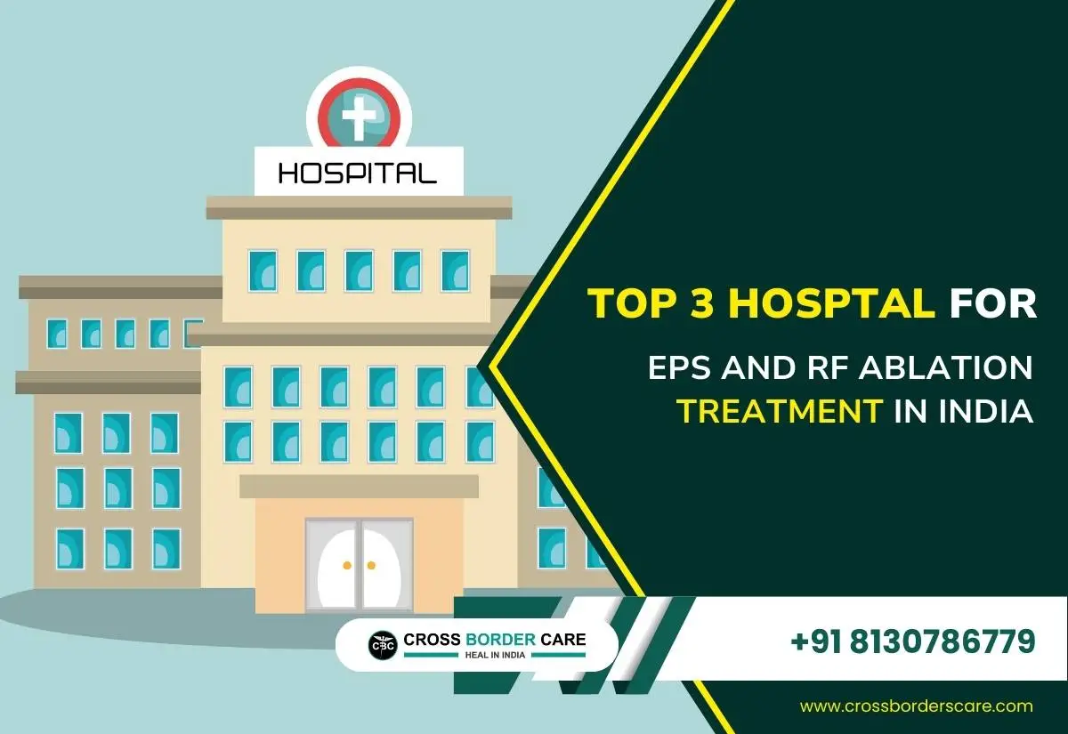
So, here we bring a well-researched and detailed list of the top and best three hospitals for EPS and RF ablation in India. It is easy to go through the key reasons for our recommendations and the exclusive features of these facilities.
Top Three Hospitals for EPS and RF Ablation in India
1. Indraprastha Apollo Hospital
First on our list of the best hospitals for EPS and RF ablation in India is the reputed Indraprastha Apollo Hospital. The exceptional medical facilities offered by this hospital are synonymous with exemplary patient care with the best combination of advanced technology and infrastructure.
Why Does Cross Border Recommend Indraprastha Apollo Hospital for EPS and RF Ablation in India?
Team of best heart surgeons
This hospital brings a team of top-notch heart surgeons for quick help to cardiac patients. Some of the top names of the cardiac team of Indraprastha Apollo Hospital cover coveted personalities like Dr. Vanita Arora, Dr. Mahesh Chandra Garg, Dr. Niranjan Hiremath, Dr. N.N. Khanna, and the list goes on.
Availability of the best heart diagnostics and treatments
This hospital is efficient in several diagnoses and treatments related to cardiac disorders. It covers EPS, RF Ablation, CRT, 3D ECHO, Doppler studies, TMT, Fetal ECHO, and others. Medical professionals have performed several processes like implanting pacemakers, coronary angioplasty, stent placement, defibrillation, heart valve surgeries, and CABG.
Offers the Apollo Edge
It was the first hospital to start the MICS techniques and systems for the effective treatment of heart patients. With a dedicated team of full-time cardiac experts, Indraprastha Apollo Hospital is capable of tackling all cardiac emergencies, complications, and complexities.
Exclusive Features of Indraprastha Apollo Hospital for EPS and RF Ablation in Delhi
| Sr. No. | Features |
| 1 | Treated several patients from Sri Lanka, Nigeria, Nepal, Ethiopia, Bangladesh, Pakistan, Afghanistan, and other locations. |
| 2 | Dedicated and fully-equipped cardiac intensive care unit. |
| 3 | Robotic-assisted cardiac surgeries for complicated cases. |
2. Fortis Escorts Heart Institute
Secondly, Fortis Escorts Heart Institute is another good option for EPS and RF Ablation in India. It is a leading heart institute armed with cutting-edge technology and clinical expertise to offer world-class treatment and personalized patient care. With the well-known cardiac professionals in its team, this hospital is respected as an eminent heart institute in Asia.
Why Does Cross Border Recommend Fortis Escorts Heart Institute for EPS and RF Ablation in India?
Best EPS and RF Ablation experts
Fortis Escorts Heart Institute offers the benefits of the top names in the electrophysiology and RF Ablation processes. It covers Dr. Anil Saxena, Dr. Aparna Jaswal, Dr. Nityanand Tripathi, and Dr. Vivek Tandon.
Offers advanced cardiac care
It offers not only EPS and RF Ablation but offers cardiac pacing, complex angioplasties, paediatric cardiology, minimally invasive cardiac surgeries, heart transplants, LVAD, TAVR etc.
Received several awards in cardiac services
Fortis Escorts Heart Institute offers a combination of skilled cardiac professionals and advanced technologies which attracted several awards and accolades. It covers the top title “Best Hospital in Cardiology.”
Exclusive Features of Fortis Escorts Heart Institute for EPS and RF Ablation in Delhi
| Sr. No. | Features |
| 1 | Center of excellence for cardiology for more than three decades. |
| 2 | Won excellence in Healthcare by AHPI for cardiac services. |
| 3 | Treated several international patients suffering from critical cardiac issues. |
3. Artemis Hospital
Last but not least, Cross Border recommends Artemis Hospital as the best hospital for EPS and RF Ablation in Delhi. The secret to the success of Artemis Heart Center lies in its team of top cardiologists, vascular surgeons, and cardiothoracic surgeons. With the availability of technology and medical expertise, Artemis Heart Center brings world-class cardiac care.
Why Does Cross Border Recommend Artemis Hospital for EPS and RF Ablation in India?
Special cardiac care diagnostic equipment and facilities
Artemis Hospital offers the best diagnostics for cardiac care covering EPS, RF Ablation, TMT, Colour Doppler, stress ECHO, 24-hour Holter monitoring, Stress Thallium test, 64-slice CT angiography, Endovascular Suite, and Flat Panel Cath Labs.
Advanced infrastructure for handling cardiac patients
This hospital offers the leading facilities for offering patient-centric cardiac treatments. It covers cardiac cath labs, endovascular operating units, dedicated cardiac operation theatres, and professional cardiac rehabilitation centres.
Offers five specialities on one platform
This hospital offers dedicated specialities under one roof only. These cover complex invasive cardiology, non-invasive cardiology, cardiothoracic and vascular surgery, paediatric cardiology, and paediatric cardiac surgeries.
Exclusive Features of Artemis Hospital for EPS and RF Ablation in Delhi
| Sr. No. | Features |
| 1 | Offers EPS and RF Ablation for SVT along with VT Ablation and AF Ablation. |
| 2 | Equipped with top cardiac cath labs, endovascular operating suits, dedicated cardiac OTs, and cardiac rehabilitation centres. |
| 3 | Specializes in non-invasive, invasive, CTVS, and paediatric heart surgeries. |
Ending Note
Cross Border encompasses a range of benefits to patients looking for EPS and RF Ablation treatment in India.
While it is easy for you to select any of the hospitals out of our carefully prepared list of the best hospitals for EPS and RF ablation in India, our experts bring much more than their guidance to you. We offer highly patient-centric treatments with a focus on savings on diagnostics and treatments.
Not to miss is the personalized follow-up in your native region along with reduced waiting time for patients in these best cardiac care units.